")
Ultrasound for Acute Pulmonary Embolus by Leanne Hartnett
Leanne Hartnett is a massive fan of bedside ultrasound. Here, she tells a story of using ultrasound for the diagnosis of acute pulmonary embolus and the decision-making process for management.
A 65-year-old man arrived at the Emergency Department with acute shortness of breath and chest tightness.
He had a background of motor neuron disease, due to which he was confined to a wheelchair. Despite this he reported a good quality of life. He enjoyed getting out and about with his wife, spending time with his family and reading the newspaper.
In saying this, he was aware of the seriousness of his disease and did not want any invasive treatments or CPR. The history and examination were unremarkable, although Leanne’s clinical suspicion of a pulmonary embolism was still high.
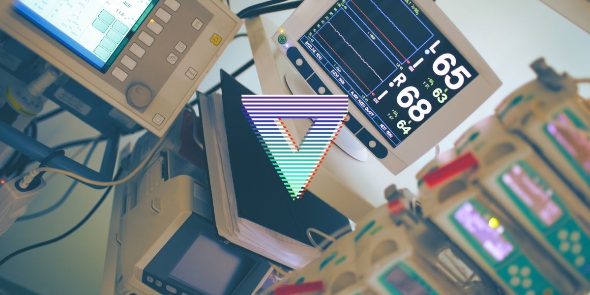
She wanted to order a CT pulmonary angiogram. However her patient was sure he would not tolerate laying flat for that length of time. So, Leanne wheeled over the ultrasound machine.
Despite the technical difficulties of the task, Leanne was able to obtain reasonable images of her patient’s cardiac structures and function. A parasternal long axis view showed a right ventricle doing not too much. A parasternal short axis view demonstrated a big right ventricle and small left ventricle.
It also demonstrated an intraventricular septum that was flattening in diastole. Finally, an apical four chamber view showed a big hyperdynamic right ventricle and something flicking about in the right atrium. It was a cord like thrombus!
She had been able to diagnose the patient using history, examination and echocardiogram alone.
Next, Leanne sought a colleague to discuss the situation and to review the literature of the different treatment modalities for right heart thrombus in transit. She suggests that the best outcomes in terms of probability of survival was with thrombolysis or embolectomy compared to anticoagulation.
Armed with the correct diagnosis and an evidence based treatment Leanne was able to successfully manage this patient. Her message is that patients who present with pulmonary embolism and right heart strain are high risk.
Thrombolysis and embolectomy are both effective strategies. Finally, basic echocardiogram skills can make a massive difference to your patients.
For more like this, head to our podcast page. #CodaPodcast